Home / See / Thinning at the crown
Presentation
Thinning at the crown
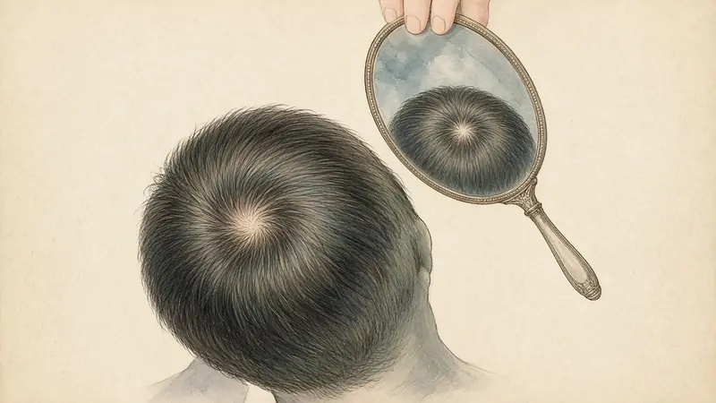
What this looks like
Thinning at the crown is a change at the vertex, the highest point of the back of the top of the head, slightly behind where a centre part usually ends. You often cannot see it yourself without two mirrors, a photograph, or a hairdresser’s comment. The hair may still look full from the front. In some people the crown is a rounder patch of show-through; in others it is simply a softer, less opaque area that only appears in overhead light.
Because the vertex is hard to self-inspect, this presentation is unusually vulnerable to a single bad photograph. A flash aimed at the crown, or wet hair combed flat, can invent a bald spot that is milder in ordinary light. That does not mean the change is imaginary. It means the first job is a fair picture, and a comparison with the back of the head, which often holds its density longer in patterned change.
What it can be confused with
| Lookalike | How it overlaps | A useful distinction |
|---|---|---|
| A Norwood vertex pattern | Norwood described a type III vertex, in which the crown thins while the hairline recession is still limited. | A clinician stages this; you can only notice that the crown and the temples may be on different schedules. |
| A Ludwig-type thinning of the top | The crown is part of the zone Ludwig graded in women. | Ludwig’s picture is usually a broader rarefaction of the top, not only a round patch. Many people sit between the drawings. |
| A cowlick or a natural crown swirl | A swirl parts itself and always shows some scalp. | Compare an older photograph. A swirl that has not changed in ten years is not a new patch. A swirl that has opened is. |
| A photograph with flash or wet hair | Both make the vertex look emptier. | Re-shoot dry, without flash, from the same height. See the photographs page. |
| A scarring patch | A rounder area of loss can be a different disease, including alopecia areata or a scarring alopecia. | A smooth, bald, or inflamed patch, especially if it appeared quickly, is not a reason to wait on more photographs. See a professional. |
How to photograph this view
Stand with your back to a window, or have someone stand behind and slightly above you. Part the hair through the crown in a straight line, or comb it as you usually wear it, but then take both versions, and keep using the same one. The camera should look down at the vertex, not toward the forehead. Include a sliver of the back of the head in the frame so that you can compare the crown with the occipital scalp later. Dry hair, no flash, no root concealer, and the same shirt colour if you can manage it: a black sweater makes every parting look wider.
Take a matching photograph of the back of the head on the same day. The front-versus-back page explains why that comparison is one of the more useful home checks you can make.
What the literature actually says
Norwood’s 1975 classification, revising Hamilton, is still the most widely used map of male pattern hair loss. It explicitly includes a type III vertex: hair loss that is primarily from the vertex, with only limited frontotemporal recession. Later types describe the crown and the frontal recession growing toward each other until the band between them is gone. Those drawings are for a patterned, progressive change. They are not a chart of every round thin area.
In women, the crown is part of the territory Ludwig graded, and it is also part of the midline-part and Christmas-tree views Olsen described. A woman can have a vertex that looks closer to a male-type pattern; later reviews of classification systems treat that as a known variant, not as a contradiction.
Clinic methods that actually count hairs at the vertex (trichoscopy, a phototrichogram, a biopsy) are not the same as a photograph, and their numbers should not be mixed with a phone picture. If a clinician has already measured the crown, let that method be the trend.
When to see a professional
A new, round, smooth, or inflamed patch should be examined, not photographed for three months. So should a crown that is changing quickly, or a vertex accompanied by pain or scaling. A dermatologist, or a trichologist listed at trichology.com, can sort a patterned crown from the lookalikes. If the picture does seem patterned, pattern support is written as support, not as a verdict, and not as a replacement for a prescribed medicine.
Which kind of support fits you?
For a gradual change at the part, crown or temples. Four one-tap questions. Nothing is stored.
A reading of a picture, not a diagnosis. Your answers stay on this page and are never stored.
Questions people actually ask
Why does my crown look worse in the evening?
Oil, flattening, and indoor overhead lights all make the vertex look less opaque. Morning photographs, in the same window light, are a kinder and more repeatable record.
If my hairline is still intact, can the crown still be a pattern?
Yes. Norwood’s type III vertex was described for exactly that combination in men, and women’s scales also allow a crown or mid-scalp change with a preserved fringe.
Keep reading
Sources
- Norwood OT. Male pattern baldness: classification and incidence. South Med J. 1975;68(11):1359–1365.
- Hamilton JB. Patterned loss of hair in man: types and incidence. Ann N Y Acad Sci. 1951;53(3):708–728.
- Ludwig E. Classification of the types of androgenetic alopecia occurring in the female sex. Br J Dermatol. 1977;97(3):247–254.
- Olsen EA. The midline part: an important physical clue to the clinical diagnosis of androgenetic alopecia in women. J Am Acad Dermatol. 1999;40(1):106–109.
- Gupta M, Mysore V. Classifications of patterned hair loss: a review. J Cutan Aesthet Surg. 2016;9(1):3–12.