Home / Measure / Comparing the top with the back
Measurement
Comparing the top with the back
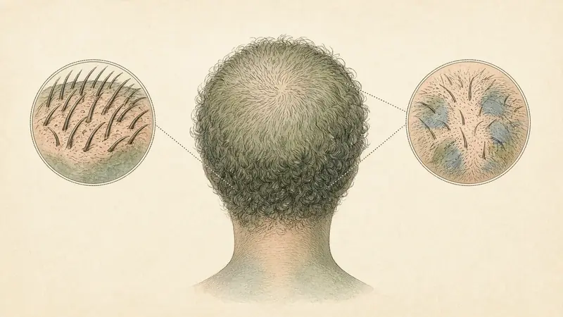
Why the back of the head is a useful control
In many patterned presentations, the sides and the back of the scalp (the occipital zone that hair-transplant surgeons call a donor area) hold their density longer than the front, the mid-line and the crown. That is not a law of nature for every person and every disease, but it is a useful contrast. A photograph of the top that looks thin, next to a photograph of the back taken on the same day in the same light, will often tell you more than a single picture of the part. A timed diffuse shed, by contrast, may thin more evenly, including at the back. The comparison does not diagnose you. It tells you which atlas pages to read next, and it is a gift to a clinician.
How to make the comparison fairly
Take the two photographs on the same day, minutes apart, with dry unstyled hair, a window, no flash, and a similar camera distance. The top view should be the one you actually care about: a mid-line part from above, or the crown from behind. The control should show the back of the head, not a new, tighter part you have just carved toward the nape to “make it fair.” If you can, include a small scale object in both frames. Repeat in three months with the same pair.
| What you might see | A cautious reading | Where to go next |
|---|---|---|
| The top looks clearly thinner than the back, and this is new | A patterned contrast is possible, including miniaturization at the front or crown. | The part, crown, or miniaturization pages, and an examination if you want it named. |
| The top and the back look similarly thinner than they used to | A more even process is possible, including a timed shed. | The diffuse-thinning page, and Hair Experts if you can name a trigger. |
| The back looks thinner than the top | Less typical for a classic androgenetic pattern, and a reason not to force one. | An examination, especially if the scalp itself looks unwell. |
| You cannot tell, because the light or the style is different | The comparison was not fair. | Re-shoot the pair under the protocol. |
What the literature actually says
Trichoscopy criteria for female androgenetic alopecia, including those proposed by Rakowska and colleagues in 2009, are built on a comparison of the frontal scalp with the occipital scalp: lower average thickness at the front, a higher share of thin hairs, and other signs read in standardized fields. That is a clinic method, but the idea (use the back as a control) is the one this home comparison borrows, without borrowing the scoring. Norwood’s map of male patterns, and the practice of harvesting transplanted hair from the occipital zone, rest on a similar observation: the back is, in many people with a pattern, relatively spared.
It is not always spared. Some diseases thin the back. Some people have more than one process. A spared back does not prove a pattern, and an unspared back does not disprove one. It is a contrast, not a verdict.
When the contrast should turn into an appointment
If the top is clearly changing relative to the back, or if you cannot make sense of the pair, be examined. A dermatologist, or a trichologist at trichology.com, can put a dermatoscope on both zones and say what a photograph can only hint at.
Questions people actually ask
I had a haircut that is shorter at the back. Can I still compare?
A much shorter back will show more scalp even if the density is unchanged. Wait until the lengths are closer, or rely on close photographs of the shafts rather than on the overall opacity.
Does a spared back mean I am a good candidate for a transplant?
That is a question for a surgeon and a clinician, not for a home photograph. This page will not have an opinion about procedures.
Keep reading
Sources
- Rakowska A, Slowinska M, Kowalska-Oledzka E, Olszewska M, Rudnicka L. Dermoscopy in female androgenic alopecia: method standardization and diagnostic criteria. Int J Trichology. 2009;1(2):123–130.
- Norwood OT. Male pattern baldness: classification and incidence. South Med J. 1975;68(11):1359–1365.
- Olsen EA. The midline part: an important physical clue to the clinical diagnosis of androgenetic alopecia in women. J Am Acad Dermatol. 1999;40(1):106–109.