Home / See / Finer, shorter hairs in the same area
Presentation
Finer, shorter hairs in the same area
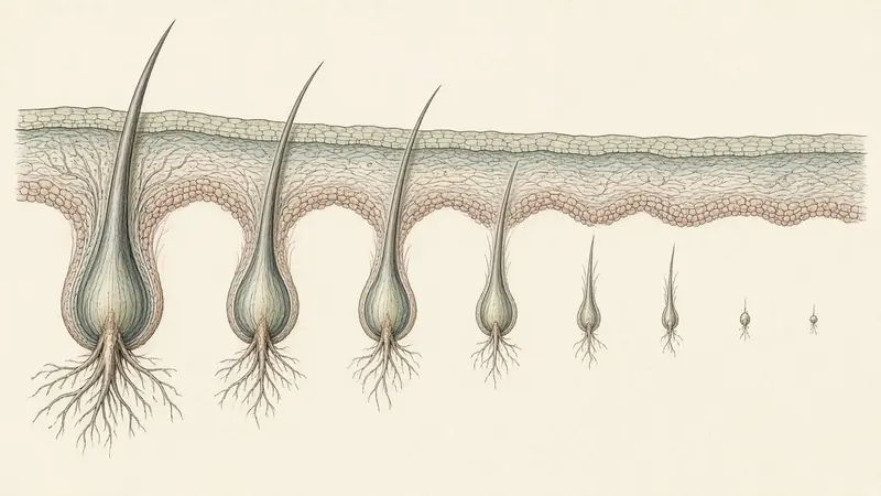
What this looks like
Miniaturization, as clinicians use the word, is a change at the follicle: hairs in an affected zone become thinner and often shorter, so that a close look at a part or a temple shows a mixture of calibers: some familiar, some finer, some almost vellus. The short hairs in that mixture are usually tapered, not snapped. The zone is often one of the patterned ones: the mid-line part, the front of the part, the crown, the temples. The back of the head, in many patterned pictures, looks more uniform.
This is not a bathroom diagnosis, and it is easy to over-call. Fine hair has always had a smaller diameter. A recent cut produces short hairs. Breakage produces short hairs of a different shape. What this page can do is tell you what a clinician is looking for, and what a careful close photograph might reasonably record, without inviting you to grade yourself on a trichoscopy scale.
What it can be confused with
| Lookalike | How it overlaps | A useful distinction |
|---|---|---|
| Naturally fine hair | Small diameter everywhere. | Miniaturization is a mix in a zone, often with a more uniform back of the head, not a lifelong fine head of hair. |
| Breakage | Short hairs in the part. | Broken ends are blunt. Miniaturized hairs are finer and tapered. See breakage versus thinning. |
| Regrowth after a shed | Short hairs at the surface. | Regrowth after a timed shed is often welcome and relatively even. A patterned mix of calibers in a typical zone is a different picture, and a clinician can help you tell them apart. |
| A self-assigned trichoscopy grade | Online checklists borrow clinic signs. | Rakowska’s criteria, for example, were written for dermoscopy in standardized fields, not for a phone camera in a bathroom. |
How to photograph this view
You want a close view of a patterned zone and a close view of the back of the head as a control, in the same light, on the same day. A window, dry hair, no flash, and a steady hand are enough to see whether the part contains a mix of thick and thin shafts. A dermatoscope attachment is optional and easy to misread; if you buy one, do not treat its pictures as a home version of a published criterion. Label the site and the date, and repeat in a few months.
What the literature actually says
Miniaturization is the follicular process behind patterned hair loss: terminal hairs, over successive cycles, spend less time growing and emerge with a smaller diameter. Trichoscopy, which is dermoscopy of the scalp, was developed in part to see that process without an immediate biopsy. In 2009, Rakowska and colleagues proposed diagnostic criteria for female androgenetic alopecia that compare the frontal scalp with the occipital scalp and that include, among other signs, a lower average hair thickness at the front and a higher share of thin hairs. Those signs were read in standardized, magnified fields. They are a clinic language.
Birch, Messenger and Messenger (2001) also matter here: many women with a clinical diagnosis of female pattern hair loss still had densities within a wide reference range. Diameter and the mix of calibers can be the earlier or the more visible story, which is why “my count is probably normal” and “my part looks different” can both be true.
When to see a professional
If you think you are seeing a mix of calibers in a patterned zone, a clinician with a dermatoscope can do in two minutes what a phone will only hint at. A dermatologist, or a trichologist at trichology.com, is the right appointment. Pattern support is written for after that kind of reading, and it will not pretend that a supplement is a microscopic examination.
Which kind of support fits you?
For a gradual change at the part, crown or temples. Four one-tap questions. Nothing is stored.
A reading of a picture, not a diagnosis. Your answers stay on this page and are never stored.
Questions people actually ask
I have always had baby hairs at my hairline. Is that miniaturization?
Not necessarily. Many hairlines have a fringe of finer hairs. A new mix of calibers in a part or at the temples, especially if it is spreading, is the change worth examining.
Can I use a cheap dermatoscope and Rakowska’s criteria at home?
You can take close pictures, and they can be useful to a clinician. Scoring published criteria yourself, from unstandardized fields, is how people over-call a diagnosis. Leave the criteria in the clinic.
Keep reading
Sources
- Rakowska A, Slowinska M, Kowalska-Oledzka E, Olszewska M, Rudnicka L. Dermoscopy in female androgenic alopecia: method standardization and diagnostic criteria. Int J Trichology. 2009;1(2):123–130.
- Birch MP, Messenger JF, Messenger AG. Hair density, hair diameter and the prevalence of female pattern hair loss. Br J Dermatol. 2001;144(2):297–304.
- Olsen EA. The midline part: an important physical clue to the clinical diagnosis of androgenetic alopecia in women. J Am Acad Dermatol. 1999;40(1):106–109.
- American Academy of Dermatology. Hair loss in women: common causes and treatment (public information).