Home / See
Presentation atlas
What you might be seeing
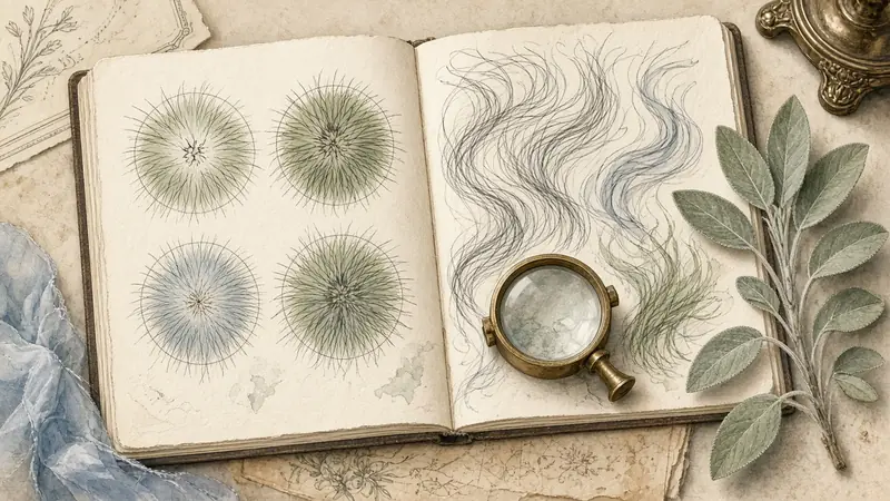
The pages in this hub are organized by the picture, not by the diagnosis. That is deliberate. A widening part, a thinner ponytail and a crown that shows in a flash photograph can share a cause, or they can come from three different ones, and you cannot tell which from a headline. What you can do, before anyone names a condition, is to look carefully and to keep a record that will still make sense in three months.
Start with the view that is closest to what you actually notice. If two views both fit, read both: the lookalike tables are there to stop a single page from becoming the whole story. If the change is timed to a birth, a medicine, an illness or a procedure, add the diffuse thinning page and then the Shed Clock on Hair Experts. If the scalp is painful, shiny, or going in patches, skip the atlas and see a professional: a dermatologist, or a trichologist listed at trichology.com.
Part
A widening midline part
A slow widening of the central part is a classic visual clue, and a lookalike for more than one condition.
Part
The Christmas tree pattern
Show-through that is widest at the front of the part and narrows as it runs back.
Crown
Thinning at the crown
A vertex change is easy to miss in a mirror and easy to over-read in one overhead photo.
Hairline
Temples moving back
Triangular recession at the temples, including in some women.
Hairline
A changing front hairline
Recession, a mature hairline, and traction can all move the front edge.
All over
Thinning that is all over
An even change, sometimes sudden, sometimes a pattern that has not declared a part yet.
Mass
A ponytail that feels smaller
A useful personal baseline that mixes density, diameter and length.
Show-through
Scalp showing through the hair
How much scalp you see depends on color, curl, light and parting, not only on how many hairs you have.
Fiber
Broken lengths vs thinning
Blunt short hairs and tapered short hairs are not the same picture.
Edges
Thinning at the edges
When the change follows the path of a tight style.
Photographs
When photos make hair look thinner
Flash, wet hair and a dark sweater can invent a change that is not there in the room.
Caliber
Finer, shorter hairs in the same area
A mix of thick and thin hairs in one view is one of the signs clinicians look for.
When the conditions change the picture
Some views are mostly about the light, the water or the camera. These pages explain how each one changes what you see, and how to check it fairly.
Light
Thinning under direct light
Overhead bulbs and bathroom spotlights make almost any part look wider.
Water
Thinning on wet hair
Wet hair clumps into strands and shows the scalp between them.
Oil
Greasy hair that looks thin
Oil groups hairs together, so the same density reads as less.
Curl
Thinning in curly hair
Curl hides show-through and shrinks length, so change is read differently.
Color
Thinning in blonde hair
Low contrast between hair and scalp changes how density looks.
Camera
Photo versus mirror
Why the phone and the mirror disagree, and which one to trust for a record.
Torn between two pictures? The comparison pages put lookalikes side by side, and the one-minute quiz walks you through the questions.
How to use this hub
Read one presentation all the way through, including the lookalikes, before you open a care page. The care pages assume you already know which picture you are dealing with. If you would rather be walked through the questions, use the Capellum Map, then come back here to the pages it names.
None of these pages is a type test for curl pattern, and none of them is a substitute for the density-and-diameter encyclopedias that already exist. When a number from a paper is useful (for example the way density changes with age in a study of women, or the way trichoscopy criteria compare the front of the scalp with the back), it is cited, and the page will say what a home check cannot do with that number.
A word about what this hub is not. It is not a quiz that issues a disease name, and it is not a gallery of before-and-after photographs. We do not use pictures of real people as if they were evidence, and we do not use generated faces as if they were patients. The plates you see are maps, still lifes, and fiber illustrations, and the record that matters is the one you make of your own scalp, under a protocol, for yourself or for a clinician.
If you arrive from a search for a single famous scale, start with the presentation that matches the view you actually have, and only then open the Norwood and Ludwig page as a vocabulary, not as a self-score. If you arrive from a bad flash photograph, start with when photos make hair look thinner and take a calmer set before you write a story about the first picture. The atlas will still be here when the set is done.
Questions people actually ask
Why not organize the site by diagnosis?
Because the reader does not have a diagnosis when they arrive, and because one picture can have more than one explanation. Organizing by presentation keeps the first job honest: look, record, and know when to get help.
Sources
- Olsen EA. The midline part: an important physical clue to the clinical diagnosis of androgenetic alopecia in women. J Am Acad Dermatol. 1999;40(1):106–109.
- Ludwig E. Classification of the types of androgenetic alopecia occurring in the female sex. Br J Dermatol. 1977;97(3):247–254.
- Norwood OT. Male pattern baldness: classification and incidence. South Med J. 1975;68(11):1359–1365.
- American Academy of Dermatology. Hair loss resource center (public information).